C O N T E N T SSee AlsoDescriptionThe DNA which carries genetic information in cells is normally packaged in the form of one or more large macromolecules called chromosomes. A chromosome, from the Greek χρώμα (color) and σώμα (body) is, minimally, a very long, continuous piece of DNA (a single DNA molecule), which contains many genes, regulatory elements and other intervening nucleotide sequences. In the chromosomes of eukaryotes, the uncondensed DNA exists in a quasi-ordered structure inside the nucleus, where it wraps around histones, and where this composite material is called chromatin. During mitosis (cell division), the chromosomes are condensed and a spindle composed of microtubules is formed. Microtubules are self-assembled from dimers of alpha and beta tubulin. Microtubules attach to chromosomes at specialized structures, the kinetochores, one of which is present on each sister chromatid. Sister chromatids are attached at an area called the centromere. This term is sometimes misleading, however, because they are not necessarily joined at the center of the chromosome. A special DNA base sequence in the region of the kinetochores provides, along with special proteins, longer-lasting attachment in this region. This is the only natural context in which individual chromosomes are visible with an optical microscope. Each chromosome has two arms, the shorter one called p arm (from the French petit, small) and the longer one q arm (q following p in the Latin alphabet). Prokaryotes? do not possess histones or nuclei. In its relaxed state, the DNA can be accessed for transcription, regulation, and replication.
Figure 1: Chromosome. (1) Chromatid. One of the two identical parts of the chromosome after S phase. (2) Centromere. The point where the two chromatids touch, and where the microtubules attach. (3) Short arm (4) Long arm. HistoryChromosomes were first observed in plant cells by Swiss botanist Karl Wilhelm von Nägeli (1817-1891) in 1842, and independently, in Ascaris worms, by the Belgian scientist Edouard Van Beneden (1846-1910). The use of basophilic aniline dyes was a fundamentally new technique for effectively staining the chromatin material inside the nucleus. Their behavior in animal (salamander) cells was later described in detail by German anatomist Walther Flemming (1843-1905), the discoverer of mitosis, in 1882. The name was invented later by another German anatomist, Heinrich von Waldeyer. In 1910, American geneticist Thomas Hunt Morgan (1866-1945) proved that chromosomes are the carriers of genes, by studying the common fruit fly. Chromosomes in plants, yeast and animalsEukaryotes (cells with nuclei such as plants, yeast, and animals) possess multiple linear chromosomes contained in the cell's nucleus. Each chromosome has one centromere, with one or two arms projecting from the centromere. The ends of the chromosomes are special structures called telomeres. DNA replication begins at many different locations on the chromosome. Chromosomes in bacteriaBacterial chromosomes are often circular but sometimes linear. Some bacteria have one chromosome, while others have a few. Bacterial DNA also exists as plasmids. The distinction between plasmids and chromosomes is poorly defined, though size and necessity are generally taken into account. Bacterial chromosomes initiate replication and one origin of replication. When linear, bacterial chromosomes tend to be tethered to the plasma membrane of the bacteria. In molecular biology application, this allows for its isolation from plasmid DNA by centrifugation of lysed bacteria and pelleting of membranes (and the attached DNA). ChromatinTwo types of chromatin can be distinguished:
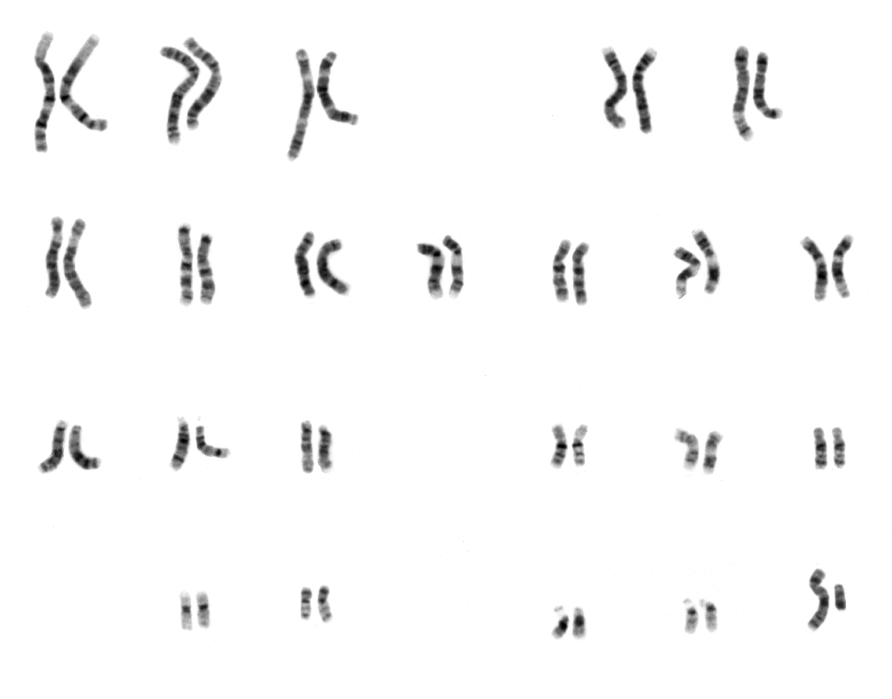
In the very early stages of mitosis, the chromatin strands become more and more condensed. They cease to function as accessible genetic material and become a compact transport form. Eventually, the two matching chromatids (condensed chromatin strands) become visible as a chromosome, linked at the centromere. Long microtubules are attached at the centromere and two opposite ends of the cell. During mitosis, the microtubules pull the chromatids apart, so that each daughter cell inherits one set of chromatids. Once the cells have divided, the chromatids are uncoiled and can function again as chromatin. In spite of their appearance, chromosomes are highly structured. For example, genes with similar functions are often kept close together in the nucleus, even if they are far apart on the chromosome. The short arm of a chromosome can be extended by a satellite chromosome that contains codes for ribosomal RNA. Number of chromosomes in different speciesNormal members of a particular species all have the same number of chromosomes. Asexually reproducing species have one set of chromosomes, which is the same in all body cells. Gametes, reproductive cells, are haploid [n] and have one set of chromosomes. Sexually reproducing species have somatic cells, body cells, which are diploid [2n] having two sets of chromosomes, one from the mother and one from the father. Gametes are produced by meiosis of a diploid germ line cell. During meiosis, the matching chromosomes of father and mother can exchange small parts of themselves (crossover), and thus create new chromosomes that are not inherited solely from either parent. When a male and a female gamete merge (fertilization), a new diploid organism is formed. Some animal and plant species are polyploid [Xn] and have more than two sets of chromosomes. Agriculturally important plants such as tobacco or wheat are often polyploid compared to their ancestral species. Wheat has a haploid number of seven chromosomes, still seen in some cultivars as well as the wild progenitors. The more common pasta and bread wheats are polyploid having 28 (tetraploid) and 42 (hexaploid) chromosomes compared to the 14 (diploid) chromosomes in the wild wheat[1]. Historical note: In 1921, Theophilus Painter claimed, based on his observations, that human sex cells had 24 pairs of chromosomes, giving humans 48 chromosomes total. It wasn't until 1955 that the number of pairs was clearly shown to be 23. KarotypeTo determine the (diploid) number of chromosomes of an organism, cells can be locked in metaphase in vitro (in a reaction vial) with colchicine. These cells are then stained (the name chromosome was given because of their ability to be stained), photographed and arranged into a karyotype (an ordered set of chromosomes), also called karyogram.
Human male karyogram. Like many sexually reproducing species, humans have special gonosomes (sex chromosomes, in contrast to autosomes for body functions). These are XX in females and XY in males. In females, one of the two X chromosomes is inactive and can be seen under a microscope as Barr bodies. Some chromosome abnormalities do not cause disease in carriers, such as translocations, or chromosomal inversions, although they may lead to a higher chance of having a child with a chromosome disorder. Abnormal numbers of chromosomes or chromosome sets, aneuploidy, may be lethal or give rise to genetic disorders. Genetic counseling is offered for families that may carry a chromosome rearrangement. Chromosomal aberrationsThe gain or loss of chromosome material can lead to a variety of genetic disorders. Examples include:
Attribution
|